18 PART I Basics of Microscopy
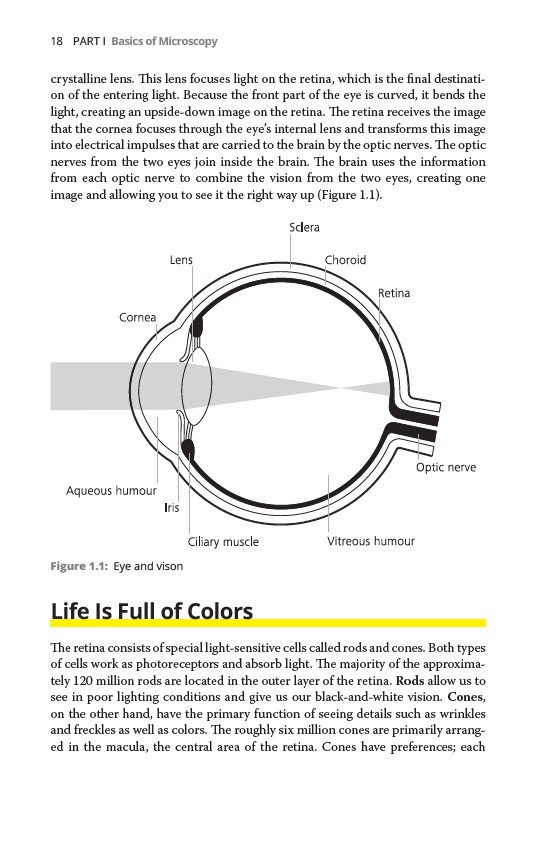
crystalline lens. This lens focuses light on the retina, which is the final destination
of the entering light. Because the front part of the eye is curved, it bends the
light, creating an upside-down image on the retina. The retina receives the image
that the cornea focuses through the eye’s internal lens and transforms this image
into electrical impulses that are carried to the brain by the optic nerves. The optic
nerves from the two eyes join inside the brain. The brain uses the information
from each optic nerve to combine the vision from the two eyes, creating one
image and allowing you to see it the right way up (Figure 1.1).
Figure 1.1: Eye and vison
Life Is Full of Colors
The retina consists of special light-sensitive cells called rods and cones. Both types
of cells work as photoreceptors and absorb light. The majority of the approximately
120 million rods are located in the outer layer of the retina. Rods allow us to
see in poor lighting conditions and give us our black-and-white vision. Cones,
on the other hand, have the primary function of seeing details such as wrinkles
and freckles as well as colors. The roughly six million cones are primarily arranged
in the macula, the central area of the retina. Cones have preferences; each